Antitumor gold nanoparticles Pr. Emmanuel Fort
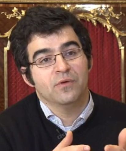

Renowned for his research into the nature and behavior of wave/matter interactions, Prof. Emmanuel Fort is the newly appointed holder of the AXA-ESPCI Chair in Biomedical Imaging at ParisTech, the Paris Institute of Science and Technology. His lab is searching for ways to use nanotechnologies to make biomedical imaging much more precise than existing methods such as MRI.
Gold nanoparticles have been known for their healing properties since ancient Greece. The odd qualities of these nanoparticles are rich in possibilities for medical research for two reasons. For one, they serve as antennae, which capture and focus waves. Then, they scatter back the energy of these waves in the form of light or convert the absorbed energy into heat. The medical challenge is to exploit these features, especially the strong interaction with light.
Part of Fort’s research uses the unique properties of these nanoparticles to image and destroy cancer tumors. When exposed to a particular wavelength of light, the particles emit light back. This enables a non-invasive, extremely high-definition biopsy, for example. What is more, because the nano-scale emitters can also produce heat, they could conceivably be used to very precisely “bake” cancerous cells, without affecting healthy ones.
Fort’s research also focuses on cellular ultramicroscopy in which live cell activity can be observed with a nanometric resolution and a sensitivity down to a single molecule.
Reaching these ultimate detection limits is most challenging but crucial to understanding the mechanisms involved in numerous diseases, such as Alzheimer's. Fort uses innovative principles to specifically improve the axial resolution and obtain 3D images of cells with a nanometer resolution. Overall, Fort’s goal is to create new types of imagery that marry diagnosis and therapy techniques into a single, unified form of treatment.

Discover research projects related to the topic
Mental Health & Neurology
Extreme Weather Events
Pollution
Alzheimer's Disease, Dementia & Neurodegenerative Diseases
Droughts & Heatwaves
Air Quality
Mécénat des Mutuelle
France
CLIMABRAIN: Impacts of Extreme Weather on the Most Vulnerable Living with Alzheimer's disease
In the context of the increase in extreme weather events due to climate change, the project led by Tarik Benmarhnia... Read more
Tarik
BENMARHNIA